Ovarian Cancer Explained
There are four stages of ovarian cancer. Each stage identifies the size and location of the tumor. The more advanced stage of ovarian cancer, the more difficult it is to treat.
This is the earliest form of ovarian cancer. Cancer is found in one or both ovaries.
ovarian cancer is divided into three stages.
Stage I:
Stage IA: Cancer is present in one ovary.
Stage IB: Cancer is present in both ovaries.
Stage IC: Cancer is present in one or both ovaries and cancer is found on the outside surface of one or both ovaries, or the outer covering of the tumor has ruptured, or cancer cells are found in the fluid or tissue linings of the abdomen.
---------------------------------------------------------------------------------------------------------------Stage II:
In stage II ovarian cancer, cancer is present in one or both ovaries, and has spread to other parts of the pelvic region. There are three stages in stage II ovarian cancer.
Stage IIA: Cancer has spread to the uterus and/or fallopian tubes.
Stage IIB: Cancer is in one or both ovaries and has spread to other organs in the pelvic region such as the bladder, rectum, or sigmoid colon.
Stage IIC: Cancer is found in one or both ovaries and has spread to the uterus, fallopian tubes, bladder, sigmoid colon, or rectum.
Cancer may also be present is tissue and fluid samples of the lining of the abdominal cavity.


DELIBERATELY DEBBIE SHARES THE MESSAGE!

|
dd_awareness_.pdf Size : 185.906 Kb Type : pdf |
Easy print-out documents
Ovarian cancer articles
|
|
ovca_file_medscape.pdf Size : 101.225 Kb Type : pdf |
|
|
oc_article.pdf Size : 232.369 Kb Type : pdf |
|
|
abdominal_OC_info.pdf Size : 81.057 Kb Type : pdf |
|
|
beatflyer.pdf Size : 170.294 Kb Type : pdf |
|
|
ovariancancer_complete.pdf Size : 126.11 Kb Type : pdf |
|
|
doc_ovca_titans-2.pdf Size : 830.528 Kb Type : pdf |
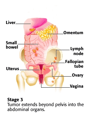
Stage III:
In Stage III Ovarian Cancer the cancer has spread outside of the pelvis into the abdominal area. This can include cancer that is found in:
3a: One or both ovaries and is grossly confined to the pelvis. Under the microscope cancer cells are found in the lining of the abdomen (peritoneum) or the omentum (a fatty layer covered by peritoneum which hangs over the intestines).
3b: One or both ovaries with confirmed implants of 2cm or less found on abdominal peritoneal surfaces.
3c: One or both ovaries with tumor growths larger than 2cm on peritoneal surfaces and/or cancer detected in lymph nodes in the upper abdomen, groin, or behind the uterus.
The majority of ovarian cancers are diagnosed at this stage.
Stage IV:
Stage IV ovarian cancer is the most advanced stage
of the disease. In this stage, cancer is found in one or both ovaries
and has spread to parts of the body beyond the abdomen, like the lungs
and liver.
-------------------------------------------------------------------------------------------------------------
How is an Ovarian Tumor Classified?
Ovarian-tumor:
Many people assume there is one simple type of ovarian tumor.This is not the case. Most forms of cancer are classified with two or more sub-types of a specific master type. When it comes to cancer in the ovaries, there are three classifications commonly identified: epithelial carcinoma, the sex cord stromal tumor, and the germ cell tumor. Each of these three classes include a series of sub-types.
Epithelial carcinoma is the more common of all forms of malignant ovarian tumors. The tumors form using cells that are located along the surface of the ovary. Adult women are at a greater risk for this form of ovarian cancer, although there are rare cases of this type of malignancy showing up in pre-teen and teenage girls.
There are several sub-types of epithelial carcinoma. Each of them has to do with the shape that the tumor takes on. A serous tumor looks somewhat like a Fallopian tube, while the mucinous tumor bears a general resemblance to a section of the GI tract. Other sub-types for the epithelial carcinoma include the endometrioid, clear cell, and transitional cell tumor.
A second class of ovarian tumor is known as the germ cell tumor. Tumors of this type are created from cells within the body of the ovary. These particular cells are actively involved in the process of egg production. While much less common that epithelial tumors, the germ cell is more likely to develop in pre-teen and teenage girls than among adult women. The chance of developing a germ cell tumor tends to diminish rapidly after the female reaches the early twenties. Even then, adult women who develop this type of tumor are more likely to find the growth is benign rather than malignant.
As with the epithelial carcinoma, the germ cell tumor is manifest in several different sub-types. Dysgerminoma is the most common sub-type, with yolk sac tumors coming in second. A less common but significant sub-type is ovarian teratoma, which has three sub-types of its own. Other forms of the germ cell tumor include choriocarcinoma and embryonal carcinoma.
The third class of the ovarian tumor is known as the sex cord stromal tumor. This particular form accounts for less than ten percent of all type of cancer found in the ovaries. Tumors of this type are created from the cells that are part of the sex chords and the gonadal stroma. In women diagnosed with some sub-type of sex cord stromal cancer, there is a significant chance of a chance in the frequency of the menstrual cycle, the appearance of more facial hair, and the development of a hoarse voice. There are several sub-types of sex cord stromal tumors;
* Sertoli-Leydig cell and
* Granulosa cell tumors are the most common. Others include;
* Fibrosarcoma
* Thecoma, and
* Sclerosing stromal tumor
Each of these forms or classes of ovarian cancer is detectable at various points in their formation. Some of them are influenced by functions involving the BRCA1, BRCA2 or HNPCC genes and may move from benign to borderline ovarian tumors before they are detected. Some serve as an example of the connection between endometriosis and ovarian cancer, or behave in a manner that is much like primary peritoneal carcinoma, a form of cancer that mimics the symptoms associated with many forms of ovarian cancer.
-------------------------------------------------------------------------------------------------------------
Ovarian Epithelial Carcinoma Explained
Epithelial carcinoma is one of the three main classes of the ovarian tumor. Reported most often in women who have reached adulthood, this form of cancer is formed from cells that are located along the surface of the ovary. Epithelial ovarian tumors are easily the most common form of cancer that is found in the ovaries, accounting for just under 60% of all diagnosed tumors and over 80% of malignant tumors associated with the ovaries.
There are five sub-types of epithelial carcinoma. Here is a short description of each:
serous-epithelial-carcinoma Serous Epithelial Ovarian Cancer
This is the most common type of ovarian cancer. In fact, when most people think in terms of ovarian cancer, this is the form they are referring to. In size, a serous tumor may be almost impossible to spot or grow to a point that the tumor fills most of the abdominal cavity.
Serous tumors are likely to be benign or borderline prior to the age of fifty. However, the chances of this type of epithelial carcinoma developing into a malignancy increase significantly among older females. If there is a history of ovarian cancer in the family line, the risks are also increased at any age.
Even among females who develop a serous tumor, the five-year survival rate is very high. If the tumor is diagnosed as benign or borderline, there is a 70-100% chance of full recovery. Depending on the severity of the malignancy, the chances of surviving for five years after the diagnosis range anywhere between 25-90%. As with any form of cancer, early detection increases the chances of survival.
mucinous-epithelial-carcinoma Mucinous Epithelial Ovarian Cancer
These tumors are another type of epithelial cancer. Less common than serous tumors, the mucinous variety still account for roughly 25% of the ovarian cancers that are identified and treated. Developing a mucinous tumor early in life is extremely rare.
Most cases involve women who are in between puberty and menopause. Even though the majority of mucinous tumors are benign, around 15% of reported cases involve a malignancy.
When it comes to survival rates, women diagnosed with mucinous tumors that are benign or borderline have up to a 95% chance of living for at least ten years after diagnosis. If there is a malignancy present, there is still a chance that two out of every three women with a mucinous tumor will live another ten years.
endometrioid-epithelial-carcinoma Endometrioid Epithelial Ovarian Cancer
Endometrioid tumors tend to be malignant in most cases. Accounting for roughly 20% of all cases of ovarian cancer, this form of ovarian epithelial carcinoma tends to develop into a shape that is somewhat similar to endometrial glands.
As the tumor grows, it will develop a series of branches that can invade surrounding tissue and organs, allowing the tumor to feed with greater ease and become more solid.
Survival after the appearance of an endometrioid tumor depends a great deal on how early the tumor is identified and treated. Because of the high possibility of malignancy, aggressive forms of treatment such as chemotherapy and possibly surgery should commence as soon as possible. If the tumor is caught in time and successfully treated, patients have a five-year survival rate of 75%.
clear-cell-epithelial-carcinoma Clear Cell Epithelial Ovarian Cancer
Clear cell tumors are a type of epithelial carcinoma that can sometimes develop in conjunction with endometrioid tumors. Containing plenty of clear cytoplasm, clear cell tumors are extremely dangerous.
Early detection and treatment increase the chances of survival after five years to 65%. However, if the tumor is not identified in its early stages, the chances of recovery are extremely low.
transitional-cell-tumor Transitional Cell Epithelial Ovarian Cancer
The Brenner tumor, also known as the transitional cell tumor, can be either extremely small or grow significantly. Tumors of this type may be solid masses or contain a fair amount of fluid. When the tumor has begun to invade surrounding tissue, there is a good chance that the growth is malignant.
All types of epithelial cancer are treatable in the early stages. Depending on the circumstances, chemotherapy may be tried as a first round of treatment, followed by surgery.
SEE FILE ON CA-125 TESTING (below)
|
|
CA-125.pdf Size : 66.43 Kb Type : pdf |

While the symptoms for ovarian cancer tend to be nonspecific and can mimic nongynecologic conditions, a large national study shows that an overwhelming majority of women diagnosed with ovarian cancer did have symptoms, sometimes even in the early stages.
The most common symptoms reported include:
A simple check-list:
* Lower abdominal and leg pain
* Sudden weight loss or gain
* Change in bowel or bladder function
* Nausea
* Swelling in the legs